First ever recording of CRISPR technology during DNA cutting
First ever recording of CRISPR technology during DNA cutting
Gene editing technology in CRISPR-Cas9 has been in use for at least a few years, but until now scientists have not directly observed the technology in action.
CRISPR (Clustered Regularly Interspaced Short Palindromic Repeats) is a system used by bacteria to defend themselves against viruses. It is such a defense mechanism, a repeated sequence of fragments in the DNA. It was noticed by Yoshizumi Ishino in the late 1980s. Last century in the genome of the bacterium Escherichia coli. CRISPR contains information about the DNA of viruses and combined with a DNA-cutting enzyme – Cas9 endonuclease provides a defense system against viruses. The technique is correctly called CRISPR-Cas9, but for simplicity the first part of the name is used.
Thanks to the gene editing technology can program genes with precision down to single nucleotide – „letters” forming genetic information. It should be added that it is a relatively simple and inexpensive technique. Thanks to CRISPR, scientists have managed to replace a defective gene in a human embryo or remove the HIV virus from the body. A spectacular example of its use was the encoding in the DNA of bacteria… kr tki film. and then reading this film, after extracting it from the DNA of successive generations of bacteria. However, the technology can lead to unwanted mutations, US researchers reported.
The CRISPR method itself, in a nutshell, works by recognizing foreign DNA through mRNA, which re responsible for transferring genetic information. The entire CRISPR sequence is then divided into kr The tighter pieces containing a viral DNA fragment and a CRISPR sequence. Based on this information, a viral transcript is created, which ry is memorized by the com rka and used in the fight against the virus. In the case of infection, the pattern of the attacking virus binds to the Cas9 enzyme and cuts the attacker into pieces completely inactivating it. The excised pieces are then added to the CRISPR sequence, a kind of threat database. Patterns can be created by humans so we can interfere with genes, substituting or cutting out dangerous fragments.
CRISPR-Cas9 activities take place on such a small scale that it is very difficult to directly observe the functioning of the technology. But difficult does not mean impossible. In a paper published in the journal „Nature Communications”, zesp ł scientist led by Mikihiro Shibata of Kanazawa University and Hiroshi Nishimasu of the University of Tokyo presented a visual observation of how CRISP-Cas9 works.
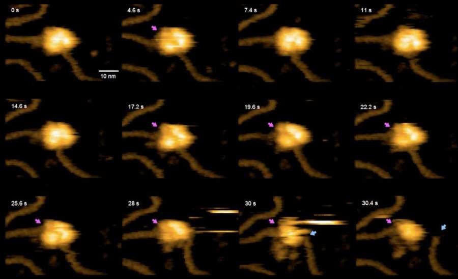
Single-molecule movie of DNA search and cleavage by CRISPR-Cas9. pic.twitter.com/3NQxmbvzJF
To an amateur, the footage will be hard to interpret, but what it shows is what many consider one of the biggest breakthroughs of the in genetics in recent times. These light brown lines are DNA strands. Ż The dot is CRISPR with the Cas9 cutting enzyme. The purple arrows indicate where the re „bites” enzyme, followed, as indicated by the gray arrow, by the separation of a severed strand of DNA.
The recording was first presented at a conference on CRISPR technology in Montana, USA. This is currently the hottest topic in genetics. – I was sitting in the front rows when the recording was played back. I heard sighs of awe behind me – said Sam Sternberg, who ry wsp He worked with the pioneer of this technology – Jennifer Doudna of the University of California at Berkeley. The scientist added that he had never encountered such a reaction at a scientific conference.
What is shown in the video is nothing new. Researchers have long known how CRISPR works. However, the evidence was always indirect. Support Modern biochemical research is a series of complex solutions that re allow inferences about the behavior of molecules too small to see. The work of Japanese scientists it changes.
The recording was made with an enhanced high-speed atomic force microscopy (AFM), which ry enables imaging of surfaces with a resolution of the order of dimension in a single atom by exploiting the forces of interatomic interactions. Standard AFM (atomic force microscopy) is used to obtain images of the in static, but its faster version allows recording of dynamic processes .